


Eman Zahran1* and Al-Zahraa Mamdouh2
1Department of Aquatic Animal Medicine, Faculty of Veterinary Medicine, Mansoura University, Mansoura, Egypt
2National Institute of Oceanography and Fisheries (NIOF), Cairo, Egypt
*Corresponding author: [email protected]
Mycotoxins, secondary metabolites produced by fungi, can significantly affect the fish gut microbiota, leading to intestinal damage, inflammation, and microbial imbalance.
![]() They alter the abundance and activity of microbial populations in the digestive tract.
They alter the abundance and activity of microbial populations in the digestive tract.
Mycotoxins such as aflatoxin B1 (AFB1) can increase the number of potentially harmful bacteria while reducing beneficial bacteria in various fish species. These effects include:
 Shifts in microbial diversity
- Changes in dominant bacterial groups
- Disruptions of the intestinal barrier
Mitigation strategies to address these effects include dietary supplements such as yeast cell wall extract, sodium butyrate, and organically modified clinoptilolite. These additives have shown promise in:
- ✓ Restoring gut bacterial diversity and composition
- ✓ Improving intestinal health
- ✓ Minimizing the negative impacts of mycotoxin exposure on the fish gut microbiota
The relationship between mycotoxins and the gut microbiota is bidirectional, with some bacteria possibly involved in detoxifying mycotoxins.
Therefore, this article focuses on how different mycotoxins affect the fish gut microbiota and explores strategies to mitigate their harmful effects.
MYCOTOXINS AND MICROBIOTA: key players in fish physiology and health
Mycotoxins are secondary metabolites produced by filamentous fungi or molds that contaminate various feedstuffs.
⇒They are mainly produced by fungal strains belonging to the genera Aspergillus, Fusarium, Penicillium, Claviceps and Alternaria (Ismaiel y Papenbrock, 2015).
To date, more than 400 different mycotoxins have been identified (Ji et al., 2016). Among them, the most detected are:
 Aflatoxins (AFs)
- Ochratoxin A (OTA)
- Patulin
- Fumonisins
- Citrinin
- Ergot alkaloids
- Trichothecenes (including deoxynivalenol (DON) and T-2 toxin (T-2))
- Zearalenone (ZEN)
- (Ismaiel and Papenbrock, 2015)
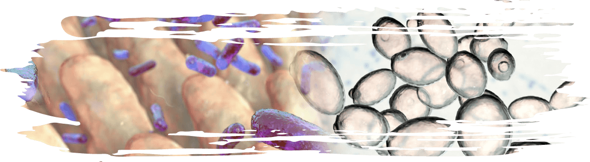
The term microbiota refers to a community of microorganisms that reside within a host and contribute to its well-being by carrying out vital functions and biological processes.
These microbial communities can be found in various human and animal organs, including the gut, oral cavity, vaginal tract, eyes, and skin (del Castillo et al., 2018).
The gut microbiota is composed of a diverse community of microorganisms—including bacteria, viruses, and fungi—that inhabit the gastrointestinal (GI) tract of living organisms (Liew and Mohd-Redzwan, 2018), playing a crucial role in supporting host health and regulating numerous physiological processes, not only within the GI tract but also in other organs and the immune system (Sekirov et al., 2010).
This microbial ecosystem can:
- ✓ Modulate the gut epithelial barrier
- ✓ Regulate inflammation
- ✓ Synthesize vitamins
- ✓ Ferment dietary fibers
- ✓ Provide protection against colonization by harmful pathogens
- (Kogut and Arsenault, 2016; Maslowski and Mackay, 2011)
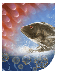
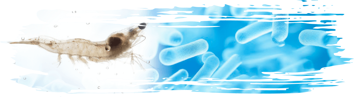
In fish, the intestinal microbiota consists of many commensal microbes whose diversity and abundance vary among species.
![]() This commensal biota plays a significant role in establishing a favorable microecological environment for fish.
This commensal biota plays a significant role in establishing a favorable microecological environment for fish.
It is essential for normal gut development and function, metabolism, immune responses, disease resistance, and the overall fish health (Li et al., 2024; Meng et al., 2023).

The effect of mycotoxins en la microbiota intestinal de los peces
Mycotoxins can cause significant harm to the fish gut microbiota, leading to a wide range of toxic effects and the disruption of gut homeostasis.
 They contribute to intestinal damage, inflammation, and microbial dysbiosis (Elmassry et al., 2022).
These toxins influence the gut microbiota by altering its abundance and activity, primarily affecting microbial populations within the digestive tract.
⇒Such changes can occur at various taxonomic levels, including species, genera, and phyla.
The modulation of the gut microbiota may result directly from the antimicrobial properties of the mycotoxins themselves or indirectly from the damage they cause to intestinal cells, which can trigger the release of antimicrobial substances (Mamdouh y Zahran).
Several experimental studies have investigated how different mycotoxins negatively affect the gut microbiota and fish intestinal health:
![]() Dietary exposure of L. maculatus to 1 mg/kg AFB1 for 56 days increased the abundance of the genera Pseudomonas, Aeromonas, and Klebsiella in the intestine, suggesting that AFB1 had an adverse effect on intestinal health, given that these bacterial genera are pathogenic.
Dietary exposure of L. maculatus to 1 mg/kg AFB1 for 56 days increased the abundance of the genera Pseudomonas, Aeromonas, and Klebsiella in the intestine, suggesting that AFB1 had an adverse effect on intestinal health, given that these bacterial genera are pathogenic.
However, this concentration did not have a significant effect on either alpha or beta diversity of the intestinal microbiota (Peng et al., 2022b).
![]() Dietary exposure of gibel carp to 50 μg/kg AFB1 for 28 days increased the abundance of Aeromonas, an opportunistic pathogenic bacterium, while decreasing the abundance of the potentially beneficial bacterium Cetobacterium, which improves digestion and produces large amounts of vitamin B.
Dietary exposure of gibel carp to 50 μg/kg AFB1 for 28 days increased the abundance of Aeromonas, an opportunistic pathogenic bacterium, while decreasing the abundance of the potentially beneficial bacterium Cetobacterium, which improves digestion and produces large amounts of vitamin B.
It also reduced the richness and diversity of bacterial communities and altered the overall intestinal bacterial composition of fish (Xue et al., 2023).
![]() Wang et al. (2018) reported that dietary exposure of Pacific white shrimp (Litopenaeus vannamei) to AFB1 at 5 ppm for 30 days increased the relative abundance of cyanobacteria.
Wang et al. (2018) reported that dietary exposure of Pacific white shrimp (Litopenaeus vannamei) to AFB1 at 5 ppm for 30 days increased the relative abundance of cyanobacteria.
A high abundance of cyanobacteria has been associated with the production of hepatotoxic microcystins, which have detrimental effects on aquatic animals (Jia et al., 2016; Kang et al., 2012).
![]() AFB1 exposure also increased the relative abundances of Vibrio and Photobacterium, both of which are recognized as pathogenic bacteria.
AFB1 exposure also increased the relative abundances of Vibrio and Photobacterium, both of which are recognized as pathogenic bacteria.
![]() Dietary AFB1 at a level of 1.0 mg/kg increased the abundance of Enterobacter (a Gram-negative opportunistic pathogen) in the intestine of e Lateolabrax maculatus (Lawley and Walker, 2013).
Dietary AFB1 at a level of 1.0 mg/kg increased the abundance of Enterobacter (a Gram-negative opportunistic pathogen) in the intestine of e Lateolabrax maculatus (Lawley and Walker, 2013).
The increased abundance of Enterobacter correlates with higher serum levels of lipopolysaccharides (LPS), which are endotoxins present in the outer membrane of Gram-negative bacteria (Eng et al., 1993).
⇒As Enterobacter becomes more prevalent in the gut, it contributes to elevated LPS levels. This increase in LPS can trigger intestinal inflammation and cause intestinal injury by disrupting the tight junctions that maintain the integrity of the intestinal barrier (Peng et al., 2022a).
![]() Small doses of AFB1 (100 μg/kg) produced slight differences in the ACE, Shannon, and, Simpson indices of alpha diversity in the intestinal microbiota of turbot. However, AFB1 altered the relative abundance of dominant bacteria; unlike in untreated fish, Bacteroidota became the dominant phylum with the highest abundance.
Small doses of AFB1 (100 μg/kg) produced slight differences in the ACE, Shannon, and, Simpson indices of alpha diversity in the intestinal microbiota of turbot. However, AFB1 altered the relative abundance of dominant bacteria; unlike in untreated fish, Bacteroidota became the dominant phylum with the highest abundance.
More importantly, AFB1 decreased the relative abundance of beneficial probiotic bacteria, including Bifidobacterium, Stenotrophomonas, Paeniclostridium, Catonella, Agathobacter, Dorea, Faecalibaculum, and Anaerostipes (Zhang et al., 2023).
![]() AFB1 at a dose of 1.0 mg/kg in the diet of largemouth bass for 56 days increased the relative abundance of Firmicutes and Mycoplasma while decreasing the abundance of Proteobacteria.
AFB1 at a dose of 1.0 mg/kg in the diet of largemouth bass for 56 days increased the relative abundance of Firmicutes and Mycoplasma while decreasing the abundance of Proteobacteria.
An increase in Firmicutes may improve the efficiency of energy absorption, but it is also associated with obesity and metabolic disorders (Sasidharan Pillai et al., 2024), which may reflect changes in host energy metabolism and increase the risk of obesity. In addition, increased Mycoplasma abundance is commonly linked to intestinal inflammation and immune disorders.
![]() A decrease in Proteobacteria abundance may reduce microbial diversity, making the intestinal ecosystem more susceptible to dysbiosis and related diseases (Fassarella et al., 2021), thereby triggering intestinal inflammation and immune dysfunction (Hou et al., 2025).
A decrease in Proteobacteria abundance may reduce microbial diversity, making the intestinal ecosystem more susceptible to dysbiosis and related diseases (Fassarella et al., 2021), thereby triggering intestinal inflammation and immune dysfunction (Hou et al., 2025).
![]() Dietary exposure of largemouth bass (Micropterus salmoides) to 44.33 ppm DON + 100 ppb AFB1 for 8 weeks decreased the relative abundance of the phylum Cyanobacteria, while exposure to 1.5 ppm DON + 20 ppb AFB1 decreased the relative abundance of Proteobacteria.
Dietary exposure of largemouth bass (Micropterus salmoides) to 44.33 ppm DON + 100 ppb AFB1 for 8 weeks decreased the relative abundance of the phylum Cyanobacteria, while exposure to 1.5 ppm DON + 20 ppb AFB1 decreased the relative abundance of Proteobacteria.
Both doses, however, increased the abundance of Fusobacteriota and Actinobacteriota. At the genus level, both doses increased the relative abundance of Cetobacterium, while the higher mycotoxin concentration also increased the abundance of Plesiomonas (Yu et al., 2025).
![]() Nile tilapia (Oreochromis niloticus) fed a diet containing AFB1 (40 μg/kg), FB (600 μg/kg), ZEN (50 μg/kg), and DON (150 μg/kg) for 42 days showed a significant increase in Proteobacteria and a significant decrease in Firmicutes (Hussein et al., 2024).
Nile tilapia (Oreochromis niloticus) fed a diet containing AFB1 (40 μg/kg), FB (600 μg/kg), ZEN (50 μg/kg), and DON (150 μg/kg) for 42 days showed a significant increase in Proteobacteria and a significant decrease in Firmicutes (Hussein et al., 2024).
A bidirectional relationship between mycotoxins and gut microbiota
Mycotoxin-induced toxicity primarily occurs through the consumption of contaminated food, making the gastrointestinal tract (GIT) the initial target of exposure.
 At the same time, the GIT also serves as the body’s first line of defense against harmful compounds.
![]() A clear example of this in fish was demonstrated by Yang et al. (2020), who reported that feeding turbot (Scophthalmus maximus) with an AFB1-contaminated diet at a dose of 500 µg/kg altered the alpha diversity of the intestinal microbiota. Specifically, OTUs, Chao1, ACE, and Shannon indices were significantly reduced.
A clear example of this in fish was demonstrated by Yang et al. (2020), who reported that feeding turbot (Scophthalmus maximus) with an AFB1-contaminated diet at a dose of 500 µg/kg altered the alpha diversity of the intestinal microbiota. Specifically, OTUs, Chao1, ACE, and Shannon indices were significantly reduced.
In addition, the relative abundance of beneficial bacteria— including Lactobacillus, Lactococcus, Streptococcus and Faecalibacterium, was significantly decreased.
These reductions were attributed to the fact that the intestinal microbiota, particularly lactic acid–producing bacteria, may be involved in the detoxification of AFB1 (Brown et al., 2011).
⇒Lactic acid has been shown to degrade AFB1 into the less toxic compounds AFB2 and AFB2a (Aiko et al., 2016).
Moreover, some strains of Lactobacillus, Lactococcus, Streptococcus, and Bifidobacterium can detoxify AFB1 directly through cell-binding mechanisms (Guan et al., 2008; Hamidi et al., 2013).
![]() In another example, an AFB1 challenge at a dose of 5 ppm in the feed of Pacific white shrimp (Litopenaeus vannamei) for 30 days increased the relative abundance of Proteobacteria and Firmicutes while decreasing the abundance of Bacteroidetes.
In another example, an AFB1 challenge at a dose of 5 ppm in the feed of Pacific white shrimp (Litopenaeus vannamei) for 30 days increased the relative abundance of Proteobacteria and Firmicutes while decreasing the abundance of Bacteroidetes.
However, some species of Firmicutes are lactic acid bacteria that have been shown to bind and remove AFB1 via surface proteins (Wang et al., 2018).
![]() Dietary exposure of hybrid grouper to 2.23 μg/kg of AFB1 reduced the abundance of Prevotella spp., which are essential for breaking down dietary fiber and producing short-chain fatty acids (SCFAs). AFB1 inhibited SCFA synthesis and disrupted lipid metabolism.
Dietary exposure of hybrid grouper to 2.23 μg/kg of AFB1 reduced the abundance of Prevotella spp., which are essential for breaking down dietary fiber and producing short-chain fatty acids (SCFAs). AFB1 inhibited SCFA synthesis and disrupted lipid metabolism.
At the same time, AFB1 exposure increased the abundance of the Prevotellaceae_NK3B31_group and Moryella. Moryella has been associated with the activation of T follicular helper (Tfh) cells and B cells during the early immune response (Yawen et al., 2022), whereas the Prevotellaceae_NK3B31_ group can produce lipopolysaccharides (LPS), potent bacterial endotoxins that trigger acute inflammation.
⇒This inflammatory response involves the release of cortisol (Singh et al., 2018; Wright et al., 2000), TNF-α, interleukin-1β (IL-1β), interferon-γ (IFN-γ), and various other cytokines and chemokines (Everhardt Queen et al., 2016; Shin and Ajuwon, 2018), thereby initiating an immune response against AFB1 (Liu et al., 2024).
Mitigation of the toxic effects of mycotoxins on fish gut microbiota
Several studies have reported that adding different food supplements to mycotoxin-contaminated diets can ameliorate their impact on the fish gut microbiota and help restore bacterial diversity and composition.
YEAST CELL WALL
Dietary yeast cell wall extract has been reported to possess mycotoxin adsorption capacity, thereby reducing the health risks associated with consuming contaminated feed and, by extension, enhancing gut microbiota performance.
![]() The addition of yeast cell wall extract at concentrations of 0.1, 0.2, and 0.4 % to a turbot diet containing 500 μg/kg AFB1 restored alpha diversity (richness, diversity, and phylogenetic diversity), as indicated by the Shannon, Simpson, ACE, and Chao1 indices of the gut microbiota in turbot (Yang et al., 2020).
The addition of yeast cell wall extract at concentrations of 0.1, 0.2, and 0.4 % to a turbot diet containing 500 μg/kg AFB1 restored alpha diversity (richness, diversity, and phylogenetic diversity), as indicated by the Shannon, Simpson, ACE, and Chao1 indices of the gut microbiota in turbot (Yang et al., 2020).
![]() Supplementation with 1000 mg/kg yeast cell wall extract combined with bacteria producing mycotoxindegrading enzymes in an AFB1-contaminated diet (100 µg/kg) for 8 weeks improved the intestinal microbiota of turbot and increased the abundance of several potential probiotics, including Faecalibacterium, Lachnoanaerobaculum, Stomatobaculum, Catonella, Delftia, and Stenotrophomonas (Zhang et al., 2023).
Supplementation with 1000 mg/kg yeast cell wall extract combined with bacteria producing mycotoxindegrading enzymes in an AFB1-contaminated diet (100 µg/kg) for 8 weeks improved the intestinal microbiota of turbot and increased the abundance of several potential probiotics, including Faecalibacterium, Lachnoanaerobaculum, Stomatobaculum, Catonella, Delftia, and Stenotrophomonas (Zhang et al., 2023).
![]() The relative abundance of phylum Proteobacteria was decreased, and the genus Mycobacterium increased after the inclusion of 0.2 % yeast cell wall extract in the feed of largemouth bass (Micropterus salmoides) contaminated with 44.33 ppm DON+100 ppb AFB1 for 8 weeks, while 0.1 % yeast cell wall extract managed to decrease the relative abundance of Plesiomonas genus.
The relative abundance of phylum Proteobacteria was decreased, and the genus Mycobacterium increased after the inclusion of 0.2 % yeast cell wall extract in the feed of largemouth bass (Micropterus salmoides) contaminated with 44.33 ppm DON+100 ppb AFB1 for 8 weeks, while 0.1 % yeast cell wall extract managed to decrease the relative abundance of Plesiomonas genus.
The addition of 0.1 % yeast cell wall extracts to feed contaminated with 1.5 ppm DON+20 ppb AFB1 markedly increased the abundance of phylum Fusobacteria and genus Cetobacterium and reduced the relative abundance of Mycobacterium (Yu et al., 2025).
SODIUM BUTYRATE
Sodium butyrate (SB) is widely used as an animal feed additive, and its role in mitigating the health effects of aflatoxins on the gut microbiota of largemouth bass has recently been investigated.
![]() Dietary supplementation with 2.0 g/kg SB for 56 days increased gut diversity and alleviated the reduction in microbiota diversity induced by AFB1 (1 mg/kg) in largemouth bass. SB supplementation significantly increased the Chao index in alpha diversity analysis, with a higher number of intestinal OTUs, and decreased the relative abundance of Firmicutes.
Dietary supplementation with 2.0 g/kg SB for 56 days increased gut diversity and alleviated the reduction in microbiota diversity induced by AFB1 (1 mg/kg) in largemouth bass. SB supplementation significantly increased the Chao index in alpha diversity analysis, with a higher number of intestinal OTUs, and decreased the relative abundance of Firmicutes.
By restoring intestinal flora balance, SB reduced the risk of metabolic disorders. It also decreased the abundance of Mycoplasma, thereby helping to reduce intestinal inflammation and improve host immune status.
Furthermore, SB supplementation increased the relative abundance of Proteobacteria, enhancing the stability of the bacterial community, supporting immunomodulatory functions, and improving overall host health (Hou et al., 2025).
ORGANICALLY MODIFIED CLINOPTILOLITE
Organically modified clinoptilolite has also been reported to mitigate the effects of mycotoxins on gut microbiota.
⇒This mineral, belonging to the heulandite (HEU) group, has a two-dimensional structure and exhibits ion-exchange capabilities in aqueous environments.
It is widely used in medicine, industry, and environmental applications, primarily for the removal of toxic pollutants from industrial wastewater and waste products.
In addition, it is used to adsorb and eliminate harmful substances, such as mycotoxins, from the digestive systems of humans and animals (Mastinu et al., 2019).
CONCLUSION
Mycotoxins, particularly aflatoxin B1 (AFB1), adversely affect the fish gut microbiota, leading to intestinal damage, inflammation, and microbial imbalance.
They alter microbial diversity, often increasing harmful bacteria while reducing beneficial ones, and compromise the integrity of the intestinal barrier.
The relationship between mycotoxins and the gut microbiota is bidirectional, as certain bacteria can contribute to mycotoxin detoxification.
Maintaining a healthy gut microbiome is therefore crucial for enhancing resistance to mycotoxins.
Mitigation strategies such as yeast cell wall extract, sodium butyrate, and organically modified clinoptilolite have shown promise in:
- ✓ Restoring gut bacterial composition
✓ Improving intestinal health - ✓ Reducing the negative impacts of mycotoxins

References
Aiko, V., Edamana, P., Mehta, A., 2016. Decomposition and detoxification of aflatoxin B1 by lactic acid. Journal of the Science of Food and Agriculture 96, 1959-1966.
Brown, C.T., Davis-Richardson, A.G., Giongo, A., Gano, K.A., Crabb, D.B., Mukherjee, N., Casella, G., Drew, J.C., Ilonen, J., Knip, M., 2011. Gut microbiome metagenomics analysis suggests a functional model for the development of autoimmunity for type 1 diabetes. PloS one 6, e25792. del Castillo, M.C.M., Valladares-García, J., Halabe-Cherem, J., 2018. Microbioma humano. Revista de la Facultad de Medicina UNAM 61, 7-19.
Elmassry, M.M., Zayed, A., Farag, M.A., 2022. Gut homeostasis and microbiota under attack: Impact of the different types of food contaminants on gut health. Critical reviews in food science and nutrition 62, 738-763.
Eng, R.H., Smith, S.M., Fan-Havard, P., Ogbara, T., 1993. Effect of antibiotics on endotoxin release from gram-negative bacteria. Diagnostic microbiology and infectious disease 16, 185-189.
Everhardt Queen, A., Moerdyk-Schauwecker, M., McKee, L.M., Leamy, L.J., Huet, Y.M., 2016. Differential expression of inflammatory cytokines and stress genes in male and female mice in response to a lipopolysaccharide challenge. PloS one 11, e0152289.
Fassarella, M., Blaak, E.E., Penders, J., Nauta, A., Smidt, H., Zoetendal, E.G., 2021. Gut microbiome stability and resilience: elucidating the response to perturbations in order to modulate gut health. Gut 70, 595-605.
Guan, S., Ji, C., Zhou, T., Li, J., Ma, Q., Niu, T., 2008. Aflatoxin B1 degradation by Stenotrophomonas maltophilia and other microbes selected using coumarin medium. International journal of molecular sciences 9, 1489-1503.
Hamidi, A., Mirnejad, R., Yahaghi, E., Behnod, V., Mirhosseini, A., Amani, S., Sattari, S., Darian, E.K., 2013. The aflatoxin B1 isolating potential of two lactic acid bacteria. Asian Pacific Journal of Tropical Biomedicine 3, 732-736.
Hou, D., Zhao, H., Peng, K., Chen, B., Hu, J., Zhu, X., Wang, G., Cao, J., Huang, W., 2025. Sodium butyrate mitigates aflatoxin B1 toxicity in largemouth bass. Aquaculture Reports 43, 102966.
Hussein, W., Cholewińska, P., Wojnarowski, K., Szeligowska, N., Hu, F., Greguła-Kania, M., Rojtinnakorn, J., Palić, D., 2024. Dietary Mycotoxins Effects on Nile Tilapia (Oreochromis niloticus) Microbiomes Can Be Mitigated with Addition of Organically Modified Clinoptilolites. Microbiology Research 15, 2232-2246.
Ismaiel, A.A., Papenbrock, J., 2015. Mycotoxins: producing fungi and mechanisms of phytotoxicity. Agriculture 5, 492-537. Ji, C., Fan, Y., Zhao, L., 2016. Review on biological degradation of mycotoxins. Animal nutrition 2, 127-133.
Jia, J., Chen, Q., Lauridsen, T.L., 2016. A systematic investigation into the environmental fate of microcystins and the potential risk: study in Lake Taihu. Toxins 8, 170.
Kang, H.-S., Sturdy, M., Krunic, A., Kim, H., Shen, Q., Swanson, S.M., Orjala, J., 2012. Minutissamides E–L, antiproliferative cyclic lipodecapeptides from the cultured freshwater cyanobacterium cf. Anabaena sp. Bioorganic & medicinal chemistry 20, 6134-6143.
Kogut, M.H., Arsenault, R.J. 2016. Gut health: The new paradigm in food animal production (Frontiers Media SA), 71.
Lawley, T.D., Walker, A.W., 2013. Intestinal colonization resistance. Immunology 138, 1-11.
Li, M., Liang, H., Yang, H., Ding, Q., Xia, R., Chen, J., Zhou, W., Yang, Y., Zhang, Z., Yao, Y., 2024. Deciphering the gut microbiome of grass carp through multi-omics approach. Microbiome 12, 2.
Liew, W.-P.-P., Mohd-Redzwan, S., 2018. Mycotoxin: Its impact on gut health and microbiota. Frontiers in cellular and infection microbiology 8, 60.
Liu, H., Xie, R., Huang, W., Yang, Y., Zhou, M., Lu, B., Li, B., Tan, B., Dong, X., 2024. Effects of dietary aflatoxin B1 on hybrid grouper (Epinephelus fuscoguttatus♀× Epinephelus lanceolatus♂) growth, intestinal health, and muscle quality. Aquaculture Nutrition 2024, 3920254.
Mamdouh, A.-Z., Zahran, E., Mycotoxins and their impact on human and animal health.
Maslowski, K.M., Mackay, C.R., 2011. Diet, gut microbiota and immune responses. Nature immunology 12, 5-9. Mastinu, A., Kumar, A., Maccarinelli, G., Bonini, S.A., Premoli, M., Aria, F., Gianoncelli, A., Memo, M., 2019. Zeolite clinoptilolite: Therapeutic virtues of an ancient mineral. Molecules 24, 1517.
Meng, D., Hao, Q., Zhang, Q., Yu, Z., Liu, S., Yang, Y., Ran, C., Zhang, Z., Zhou, Z., 2023. A compound of paraprobiotic and postbiotic derived from autochthonous microorganisms improved growth performance, epidermal mucus, liver and gut health and gut microbiota of common carp (Cyprinus carpio). Aquaculture 570, 739378.
Peng, K., Chen, B., Sun, Y., Chen, X., Wang, Y., Huang, W., 2022a. Response of growth performance, serum metabolites, intestinal tight junction structure and bacterial microbiomes to the long-term intervention of aflatoxin B1 in Lateolabrax maculatus diets. Aquaculture Reports 22, 101005.
Peng, K., Chen, B., Wang, Y., Zhao, H., Zheng, C., Chen, X., Huang, W., 2022b. Condensed tannins protect against aflatoxin B1-induced toxicity in Lateolabrax maculatus by restoring intestinal integrity and regulating bacterial microbiota. Aquaculture 555, 738255.
Sasidharan Pillai, S., Gagnon, C.A., Foster, C., Ashraf, A.P., 2024. Exploring the gut microbiota: key insights into its role in obesity, metabolic syndrome, and type 2 diabetes. The Journal of Clinical Endocrinology & Metabolism 109, 2709-2719.
Sekirov, I., Russell, S.L., Antunes, L.C.M., Finlay, B.B., 2010. Gut microbiota in health and disease. Physiological reviews. Shin, S., Ajuwon, K.M., 2018. Lipopolysaccharide alters thermogenic and inflammatory genes in white adipose tissue in mice fed diets with distinct 18‐carbon fatty‐acid composition. Lipids 53, 885-896.
Singh, S., Natesan, R., Sharma, N., Singh, M., Rahal, A., 2018. Lipopolysaccharide exposure modifies salivary and circulating level of cortisol in goats. Small Ruminant Research 162, 30-33.
Wang, Y., Wang, B., Liu, M., Jiang, K., Wang, M., Wang, L., 2018. Aflatoxin B1 (AFB1) induced dysregulation of intestinal microbiota and damage of antioxidant system in pacific white shrimp (Litopenaeus vannamei). Aquaculture 495, 940-947.
Wright, K.J., Balaji, R., Hill, C., Dritz, S., Knoppel, E., Minton, J., 2000. Integrated adrenal, somatotropic, and immune responses of growing pigs to treatment with lipopolysaccharide. Journal of Animal Science 78, 1892-1899.
Xue, M., Fu, M., Zhang, M., Xu, C., Meng, Y., Jiang, N., Li, Y., Liu, W., Fan, Y., Zhou, Y., 2023. Aflatoxin B1 induced oxidative stress and gut microbiota disorder to increase the infection of cyprinid Herpesvirus 2 in Gibel Carp (Carassius auratus gibelio). Antioxidants 12, 306.
Yang, J., Wang, T., Lin, G., Li, M., Zhu, R., Yiannikouris, A., Zhang, Y., Mai, K., 2020. The assessment of diet contaminated with aflatoxin B1 in juvenile turbot (Scophthalmus maximus) and the evaluation of the efficacy of mitigation of a yeast cell wall extract. Toxins 12, 597.
Yawen, Z., Xiangyun, C., Binyou, L., Xingchen, Y., Taiping, L., Xuedong, Z., Jiyao, L., Lei, C., Wenyue, X., Biao, R., 2022. The dynamic landscape of parasitemia dependent intestinal microbiota shifting and the correlated gut transcriptome during Plasmodium yoelii infection. Microbiological Research 258, 126994.
Yu, Z., Zhang, Z., Teame, T., Guan, L., Wang, R., Zhu, R., Zhang, Q., Yang, H., Cui, N., Huang, Y., 2025. Yeast cell wall extract as a strategy to mitigate the effects of aflatoxin B1 and deoxynivalenol on liver and intestinal health, and gut microbiota of largemouth bass (Micropterus salmoides). Aquaculture 597, 741917.
Zhang, Z., Zhou, Y., Yang, J., Zhang, W., Mai, K., Zhang, Y., 2023. Evaluation of the effects of dietary mycotoxin-degrading adsorbent on juvenile turbot (Scophthalmus maximus L.) fed aflatoxin B1-contaminated diets. Aquaculture reports 30, 101539.
 Micotoxicosis prevention